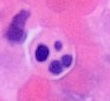
Karyorrhexis

Karyorrhexis (from Greek κάρυον karyon 'kernel, seed, nucleus' and ῥῆξις rhexis 'bursting') is the destructive fragmentation of the nucleus of a dying cell[1] whereby its chromatin is distributed irregularly throughout the cytoplasm.[2] It is usually preceded by pyknosis and can occur as a result of either programmed cell death (apoptosis), cellular senescence, or necrosis. [citation needed]
In apoptosis, the cleavage of DNA is done by Ca2+ and Mg2+ -dependent endonucleases. [citation needed]
-
Morphological characteristics of pyknosis and other forms of nuclear destruction.
-
Microscopy of an apoptotic neutrophil with nuclear fragmentation (H&E stain)
Overview
[edit]During apoptosis, a cell goes through a series of steps as it eventually breaks down into apoptotic bodies, which undergo phagocytosis. In the context of karyorrhexis, these steps are, in chronological order, pyknosis (the irreversible condensation of chromatin), karyorrhexis (fragmentation of the nucleus and condensed DNA) and karyolysis (dissolution of the chromatin due to endonucleases).
Karyorrhexis involves the breakdown of the nuclear envelope and the fragmentation of condensed chromatin due to endonucleases. In cases of apoptosis, karyorrhexis ensures that nuclear fragments are quickly removed by phagocytes. In necrosis, however, this step fails to progress in an orderly manner, leaving behind fragmented cellular debris, further contributing to tissue damage and inflammation.[3]
Process of Nuclear Envelope Dissolution During Karyorrhexis
[edit]In the intrinsic pathway of apoptosis, environmental factors such as oxidative stress signal pro-apoptotic members of the Bcl-2 protein family to eventually break the outer membrane of the mitochondria.[4] This causes cytochrome c to leak into the cytoplasm, which causes a cascade of events that eventually leads to the activation of several caspases.[4] One of these caspases, caspase-6, is known to cleave nuclear lamina proteins such as lamin A/C, which hold the nuclear envelope together, thereby aiding in the dissolution of the nuclear envelope.[5]
Process of Condensed Chromatin Fragmentation During Karyorrhexis
[edit]In the process of karyorrhexis through apoptosis, DNA is fragmented in an orderly manner by endonucleases such as caspase-activated DNase and discrete nucleosomal units are formed.[6] This is because the DNA has already been condensed during pyknosis, meaning it has been wrapped around histones in an organized manner, with around 180 base pairs per histone. The fragmented chromatin observed during karyorrhexis is made when activated endonucleases cleave the DNA in between the histones, resulting in orderly, discrete nucleosomal units.[7] These short DNA fragments left by the endonucleases can be identified on an agar gel during electrophoresis due to their unique “laddered” appearance, allowing researchers to better identify cell death through apoptosis.[8]
Nucleus Degradation in Other Forms of Cell Death
[edit]Karyorrhexis is associated with a controlled breakdown of the nuclear envelope, typically by caspases that destroy lamins during apoptosis. However, for other forms of cell death that are less controlled than apoptosis, such as necrosis (unprogrammed cell death), the degradation of the nucleus is caused by other factors. Unlike apoptosis, necrosis cells are characterized by having a ruptured plasma membrane, no association with the activation of caspases, and typically invoking an inflammatory response.[3] Because necrosis is a caspase-independent process, the nucleus may stay intact during early stages of cell death before being ripped open due to osmotic stress and other factors associated with having a hole in the plasma membrane. A specialized form of necrosis, called necroptosis, has a slightly more controlled degradation of the nucleus. This process is dependent on calpain, which is a protease that also degrades lamins, destabilizing the structure of the nucleus.[3] However, similar to necrosis, this process also involves a ruptured plasma membrane, which contributes to the uncontrolled degradation of the nuclear envelope.
Unlike karyorrhexis in apoptosis which produces apoptotic bodies to be digested through phagocytosis, karyorrhexis in necroptosis leads to the expulsion of cell contents into extracellular space to be digested through pinocytosis.[9]
Triggers and Mechanisms
[edit]The process of apoptosis, and thereby nucleus degradation through karyorrhexis, is invoked by various physiological and pathological stimuli. DNA damage, oxidative stress, hypoxia, and infections can initiate signaling cascades leading to nuclear degradation through the intrinsic pathway of apoptosis. The intrinsic pathway can also be induced through ethanol, which activates apoptosis-related proteins such as BAX and caspases.[10] Additionally, if the death receptors on a cell’s surface are activated, such as CD95, the activation of caspases and nuclear envelope degradation can be triggered as well.[5] In all of these processes, caspases such as caspase-3 play a key role by cleaving nuclear lamins and promoting chromatin fragmentation.[3] In necrosis, uncontrolled calcium influx and activation of proteases such as calpains accelerate the process, highlighting the contrasting regulatory mechanisms between necrotic and apoptotic karyorrhexis.[11]
The level of DNA damage determines whether a cell undergoes apoptosis or cell senescence. Cellular senescence refers to the cessation of the cell cycle and thus cell division, which can be observed after a fixed amount (approximately 50) of doublings in primary cells.[12] One cause of cellular senescence is DNA damage through the shortening of telomeres. This causes a DNA damage response (DDR), which, if prolonged over a long period of time, activates ATR and ATM damage kinases. These kinases activate two more kinases, Chk1 and Chk2 kinases, which can alter the cell in a few different ways. One of these ways is by activating a transcription factor known as p53. If the level of DNA damage is mild, the p53 will opt to activate CIP, which inhibits CDKs, arresting the cell cycle. However, if the level of DNA damage is severe enough, p53 can trigger apoptotic pathways which lead to the dissolution of the nuclear envelope through karyorrhexis.[13]
Pathological Implications
[edit]Karyorrhexis is a prominent feature in conditions related to cell death, such as ischemia and neurodegenerative disorders. It has been observed during myocardial infarction and brain stroke, indicating its contribution to cell death in acute stress responses.[14] Moreover, disorders such as placental vascular malperfusion have highlighted the role of karyorrhexis in fetal demise, particularly when it disrupts normal tissue homeostasis.[15]
In cancer, apoptotic karyorrhexis plays a dual role. While it facilitates controlled cell death, aiding in tumor suppression, resistance to apoptosis in cancer cells results in evasion of this pathway, promoting malignancy. Therapeutic interventions targeting apoptotic pathways attempt to restore this phase of nuclear degradation to induce tumor regression.[16]
See also
[edit]References
[edit]- ^ Zamzami N, Kroemer G (1999). "Apoptosis: Condensed matter in cell death". Nature. 401 (127): 127–8. Bibcode:1999Natur.401..127Z. doi:10.1038/43591. PMID 10490018. S2CID 36673000.
- ^ Advances in Mutagenesis Research. Springer Science & Business Media. 2012. p. 11. ISBN 9783642781933. Retrieved 11 November 2017.
- ^ a b c d Nikoletopoulou, Vassiliki; Markaki, Maria; Palikaras, Konstantinos; Tavernarakis, Nektarios (December 2013). "Crosstalk between apoptosis, necrosis and autophagy". Biochimica et Biophysica Acta (BBA) - Molecular Cell Research. 1833 (12): 3448–3459. doi:10.1016/j.bbamcr.2013.06.001. PMID 23770045.
- ^ a b Kaloni, Deeksha; Diepstraten, Sarah T.; Strasser, Andreas; Kelly, Gemma L. (2023-02-01). "BCL-2 protein family: attractive targets for cancer therapy". Apoptosis. 28 (1): 20–38. doi:10.1007/s10495-022-01780-7. ISSN 1573-675X. PMC 9950219. PMID 36342579.
- ^ a b Lindenboim, Liora; Zohar, Hila; Worman, Howard J.; Stein, Reuven (2020-04-27). "The nuclear envelope: target and mediator of the apoptotic process". Cell Death Discovery. 6 (1): 29. doi:10.1038/s41420-020-0256-5. ISSN 2058-7716. PMC 7184752. PMID 32351716.
- ^ Nagata, Shigekazu (2000-04-10). "Apoptotic DNA Fragmentation". Experimental Cell Research. 256 (1): 12–18. doi:10.1006/excr.2000.4834. ISSN 0014-4827. PMID 10739646.
- ^ Arends, M. J.; Morris, R. G.; Wyllie, A. H. (March 1990). "Apoptosis. The role of the endonuclease". The American Journal of Pathology. 136 (3): 593–608. ISSN 0002-9440. PMC 1877493. PMID 2156431.
- ^ Gong, J. P.; Traganos, F.; Darzynkiewicz, Z. (1994-05-01). "A Selective Procedure for DNA Extraction from Apoptotic Cells Applicable for Gel Electrophoresis and Flow Cytometry". Analytical Biochemistry. 218 (2): 314–319. doi:10.1006/abio.1994.1184. ISSN 0003-2697. PMID 8074286.
- ^ Wu, Ying; Dong, Guoqiang; Sheng, Chunquan (September 2020). "Targeting necroptosis in anticancer therapy: mechanisms and modulators". Acta Pharmaceutica Sinica B. 10 (9): 1601–1618. doi:10.1016/j.apsb.2020.01.007. PMC 7563021. PMID 33088682.
- ^ Fernández-Solà, Joaquim (February 2020). "The Effects of Ethanol on the Heart: Alcoholic Cardiomyopathy". Nutrients. 12 (2): 572. doi:10.3390/nu12020572. ISSN 2072-6643. PMC 7071520. PMID 32098364.
- ^ Priante, Giovanna; Gianesello, Lisa; Ceol, Monica; Del Prete, Dorella; Anglani, Franca (January 2019). "Cell Death in the Kidney". International Journal of Molecular Sciences. 20 (14): 3598. doi:10.3390/ijms20143598. ISSN 1422-0067. PMC 6679187. PMID 31340541.
- ^ Hayflick, L.; Moorhead, P. S. (1961-12-01). "The serial cultivation of human diploid cell strains". Experimental Cell Research. 25 (3): 585–621. doi:10.1016/0014-4827(61)90192-6. ISSN 0014-4827. PMID 13905658.
- ^ Surova, O.; Zhivotovsky, B. (August 2013). "Various modes of cell death induced by DNA damage". Oncogene. 32 (33): 3789–3797. doi:10.1038/onc.2012.556. ISSN 1476-5594. PMID 23208502.
- ^ Zhang, Dongjian; Jiang, Cuihua; Feng, Yuanbo; Ni, Yicheng; Zhang, Jian (2020-07-02). "Molecular imaging of myocardial necrosis: an updated mini-review". Journal of Drug Targeting. 28 (6): 565–573. doi:10.1080/1061186X.2020.1725769. ISSN 1061-186X. PMID 32037899.
- ^ Stanek, Jerzy; Drach, Alex (2022). "Placental CD34 immunohistochemistry in fetal vascular malperfusion in stillbirth". Journal of Obstetrics and Gynaecology Research. 48 (3): 719–728. doi:10.1111/jog.15169. ISSN 1447-0756. PMID 35092332.
- ^ Wong, Rebecca SY (2011-09-26). "Apoptosis in cancer: from pathogenesis to treatment". Journal of Experimental & Clinical Cancer Research. 30 (1): 87. doi:10.1186/1756-9966-30-87. ISSN 1756-9966. PMC 3197541. PMID 21943236.


