Lysosome: Difference between revisions
Appearance
Content deleted Content added
m Reverted edits by 65.12.51.212 (HG) |
No edit summary |
||
| Line 1: | Line 1: | ||
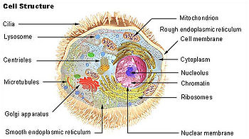
[[Image:Illu cell structure.jpg|thumb|350px|Various [[organelles]] labeled. The '''lysosome''' is labeled in the upper left.]] |
nhfhjgfhgf[[Image:Illu cell structure.jpg|thumb|350px|Various [[organelles]] labeled. The '''lysosome''' is labeled in the upper left.]] |
||
jgd |
|||
[[image:biological_cell.svg|thumb|350px|Schematic of typical animal cell, showing subcellular components. [[Organelle]]s:<br/> |
[[image:biological_cell.svg|thumb|350px|Schematic of typical animal cell, showing subcellular components. [[Organelle]]s:<br/> |
||
hi jskasdjkf;asklhnv;asje;klfjskvhjnaxkjnv;alisdijgflkxjcvnl;k jxdl;kfjgvlfjksjaloieurjmjsdfhbnmsdlk;fjg;sihjlkjnklghlksfjghio;rjftgkln dfgjkld itju dgfd df kjfdurh jklcfhj fihyujrl kfklb ;rlfgjklfcbuery98273j djh;jkdehgk;dfgfhj |
|||
(1) [[nucleolus]]<br/> |
|||
| ⚫ | |||
(2) [[cell nucleus|nucleus]]<br/> |
|||
(3) [[ribosomes]] (little dots)<br/> |
|||
(4) [[vesicle (biology)|vesicle]]<br/> |
|||
(5) rough [[endoplasmic reticulum]] (ER)<br/> |
|||
(6) [[Golgi apparatus]]<br/> |
|||
(7) [[Cytoskeleton]]<br/> |
|||
(8) smooth [[endoplasmic reticulum]]<br/> |
|||
(9) [[mitochondrion|mitochondria]]<br/> |
|||
(10) [[vacuole]]<br/> |
|||
(11) [[cytoplasm]]<br/> |
|||
(12) [[lysosome]]<br/> |
|||
(13) [[centriole]]s within [[centrosome]]]] |
|||
'''Lysosomes''' are [[organelle]]s that contain [[digestive enzyme]]s (acid [[hydrolase]]s). They digest excess or worn-out [[organelle]]s, food particles, and engulfed [[virus]]es or [[bacteria]]. The [[biological membrane|membrane]] surrounding a lysosome allows the [[digestive enzyme]]s to work at the 4.5 [[pH]] they require. Lysosomes fuse with [[vacuole]]s and dispense their enzymes into the [[vacuole]]s, digesting their contents. They are created by the addition of hydrolytic enzymes to early endosomes from the [[Golgi apparatus]]. The name ''lysosome'' derives from the Greek words ''lysis'', which means dissolution or destruction, and ''soma'', which means body. They are frequently nicknamed "suicide-bags" or "suicide-sacs" by cell biologists due to their role in [[autolysis]]. Lysosomes were discovered by the Belgian cytologist [[Christian de Duve]] in 1949. |
|||
At [[pH]] 4.8, the interior of the lysosomes is more acidic than the [[cytosol]] (pH 7.2). The lysosome's single [[Cell membrane|membrane]] stabilizes the low pH by pumping in [[proton]]s (H<sup>+</sup>) from the cytosol via [[proton pump]]s and chloride [[ion channel]]s. The membrane also protects the cytosol, and therefore the rest of the [[cell (biology)|cell]], from the [[degradative enzyme]]s within the lysosome. For this reason, should a lysosome's acid [[hydrolases]] leak into the cytosol, their potential to damage the cell will be reduced, because they will not be at their optimum pH. |
|||
==Enzymes== |
|||
Some important enzymes in these are: |
|||
*[[Lipase]], which digests [[lipid]]s |
|||
*[[Carbohydrase]]s, which digest [[carbohydrate]]s (e.g., sugars) |
|||
*[[Protease]]s, which digest [[protein]]s |
|||
*[[Nuclease]]s, which digest [[nucleic acid]]s |
|||
*[[phosphoric acid]] monoesters. |
|||
Lysosomal enzymes are synthesized in the cytosol and the [[endoplasmic reticulum]], where they receive a [[mannose|mannose-6-phosphate]] tag that targets them for the lysosome. Aberrant lysosomal targeting causes [[inclusion-cell disease]], whereby enzymes do not properly reach the lysosome, resulting in accumulation of waste within these organelles. |
|||
==Functions== |
|||
The lysosomes are used for the digestion of [[macromolecule]]s from [[phagocytosis]] (ingestion of other dying cells or larger extracellular material), [[endocytosis]] (where [[Receptor (biochemistry)|receptor protein]]s are recycled from the cell surface), and [[autophagy]] (wherein old or unneeded organelles or proteins, or microbes that have invaded the cytoplasm are delivered to the lysosome). Autophagy may also lead to [[autophagy|autophagic cell death]], a form of [[programmed cell death|programmed self-destruction]], or [[autolysis]], of the cell, which means that the cell is digesting itself. |
|||
Other functions include digesting foreign bacteria (or other forms of waste) that invade a cell and helping repair damage to the [[plasma membrane]] by serving as a membrane patch, sealing the wound. In the past, lysosomes were thought to kill cells that were no longer wanted, such as those in the tails of [[tadpole]]s or in the web from the fingers of a 3- to 6-month-old [[fetus]]. While lysosomes digest some materials in this process, it is actually accomplished through programmed cell death, called [[apoptosis]].<ref>[http://users.rcn.com/jkimball.ma.ultranet/BiologyPages/L/Lysosomes.html Lysosomes and Peroxisomes<!-- Bot generated title -->]</ref><ref>Mader, Sylvia. (2007). Biology 9th ed. McGraw Hill. New York. ISBN 978-0072464634</ref> |
|||
==Clinical relevance== |
|||
There are a number of illnesses that are caused by the malfunction of the lysosomes or one of their digestive proteins, e.g., [[Tay-Sachs disease]], or [[Pompe's disease]]. These are caused by a defective or missing digestive protein, which leads to the accumulation of substrates within the cell, impairing [[metabolism]]. |
|||
In the broad sense, these can be classified as [[mucopolysaccharidosis|mucopolysaccharidoses]], [[GM2 gangliosidosis|GM<sub>2</sub> gangliosidoses]], [[lipid storage disorder]]s, [[glycoproteinosis|glycoproteinoses]], [[mucolipidosis|mucolipidoses]], or [[leukodystrophy|leukodystrophies]]. |
|||
==Additional images== |
|||
<gallery> |
|||
Image:Localisations02eng.jpg|Proteins in different [[cellular compartment]]s and structures tagged with [[green fluorescent protein]]. |
|||
</gallery> |
|||
| ⚫ | |||
* [http://opm.phar.umich.edu/localization.php?localization=Lysosome%20membrane 3D structures of proteins associated with lysosome membrane] |
* [http://opm.phar.umich.edu/localization.php?localization=Lysosome%20membrane 3D structures of proteins associated with lysosome membrane] |
||
Revision as of 01:33, 8 October 2008
nhfhjgfhgf

jgd
[[image:biological_cell.svg|thumb|350px|Schematic of typical animal cell, showing subcellular components. Organelles:
hi jskasdjkf;asklhnv;asje;klfjskvhjnaxkjnv;alisdijgflkxjcvnl;k jxdl;kfjgvlfjksjaloieurjmjsdfhbnmsdlk;fjg;sihjlkjnklghlksfjghio;rjftgkln dfgjkld itju dgfd df kjfdurh jklcfhj fihyujrl kfklb ;rlfgjklfcbuery98273j djh;jkdehgk;dfgfhj
hghfjgjhjggb==External links=mng=
References
 This article incorporates public domain material from Science Primer. NCBI. Archived from the original on 2009-12-08.
